南湖新闻网讯(通讯员 施雪萍)近日,我校园艺林学学院植物维管生物学实验室在Plant Physiology在线发表了题为“Toward scalable wood anatomy: a toolkit for automated xylem cell identification and quantification in woody angiosperms”的研究论文。本研究构建了一个包含超过17万个精细标注实例的大规模杨树木质部图像数据集,并在此基础上对多种主流深度学习模型进行了系统评估。最终,研究开发了一套综合性智能分析工具包——PLXY-AI。该工具通过创新性提出“先分割后检测(Segmentation-then-Detection)”策略,实现了对木本被子植物横切面与弦切面中多种细胞类型的高通量、全自动识别与精确形态测量,为系统性解析林木维管组织结构与功能提供了强有力的技术支撑。
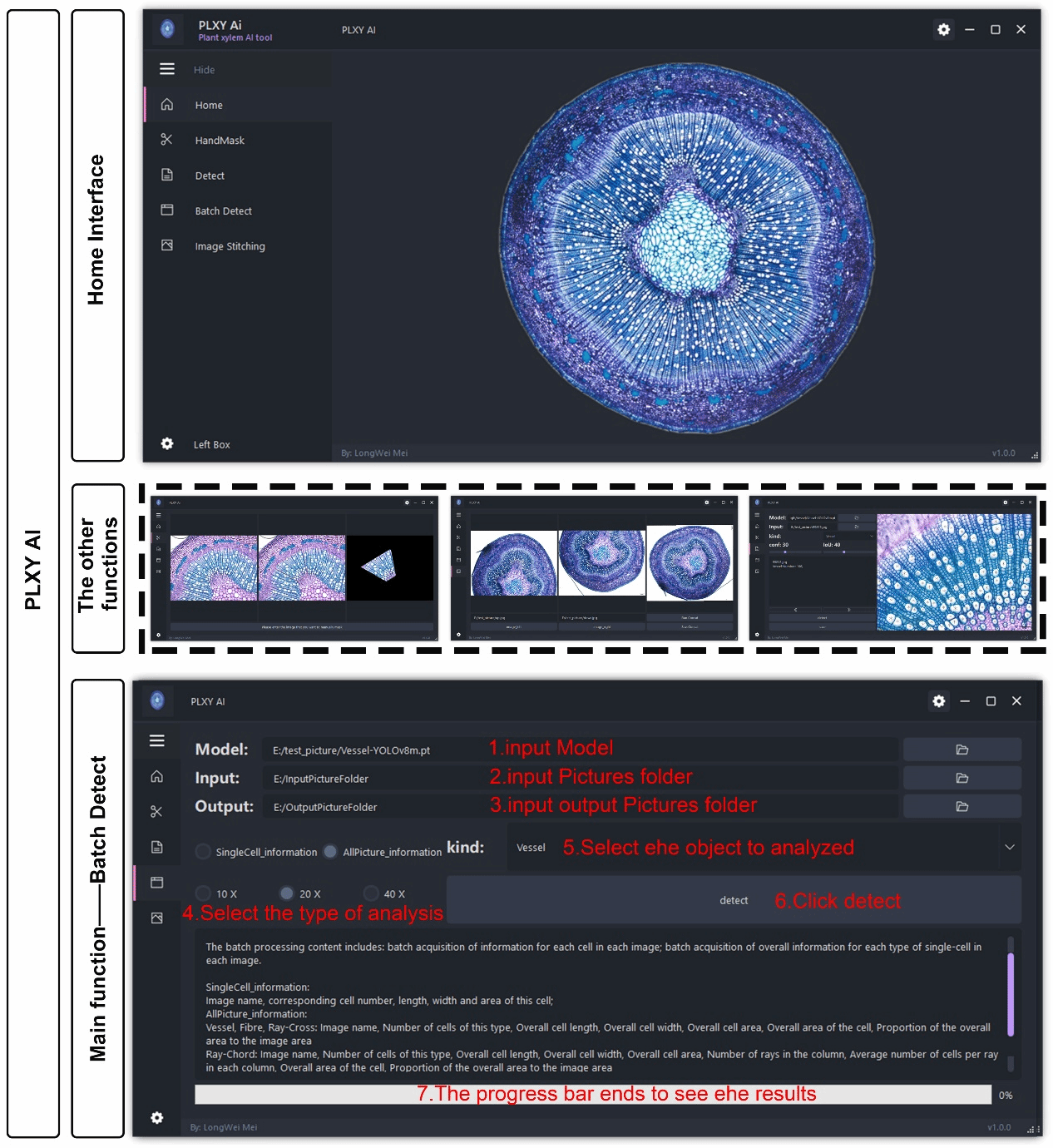
PLXY-AI 工具的用户界面
木质部(xylem)是木本植物中承担长距离水分与矿物质运输、养分再分配以及机械支撑功能的核心组织。其细胞组成(如导管、纤维和射线实质细胞)及其空间排列方式,直接决定了树木的水力传导效率、结构稳定性和环境适应能力。在木材解剖学研究中,定量木材解剖(Quantitative Wood Anatomy,QWA)是揭示细胞结构与功能关系的关键手段。然而,传统分析方法高度依赖显微镜观察和人工测量,不仅耗时费力,且易受主观因素影响而产生偏差。尽管近年来已开发出WinCELL、ROXAS等半自动化分析工具,但在处理背景复杂(如韧皮部或形成层干扰)或大规模异质样本时,其分析效率和准确性仍存在明显局限。
随着人工智能技术的快速发展,利用深度学习实现木质部细胞的自动识别与多属性定量分析,已成为木材科学和林木育种领域的迫切需求。针对上述挑战,研究通过构建杨树木质部大规模高质量数据集并开展模型系统评估,提出并验证“先分割后检测”策略在提升识别精度方面的优势,开发集成化软件工具包PLXY-AI,系统评估模型的跨物种泛化能力并建立批量化、自动化的木材解剖学分析工作流,最终形成了一套用户友好、性能稳定且可扩展的木质部细胞自动识别与多属性定量分析解决方案,为木材解剖学研究中的高通量分析提供了坚实的技术基础。
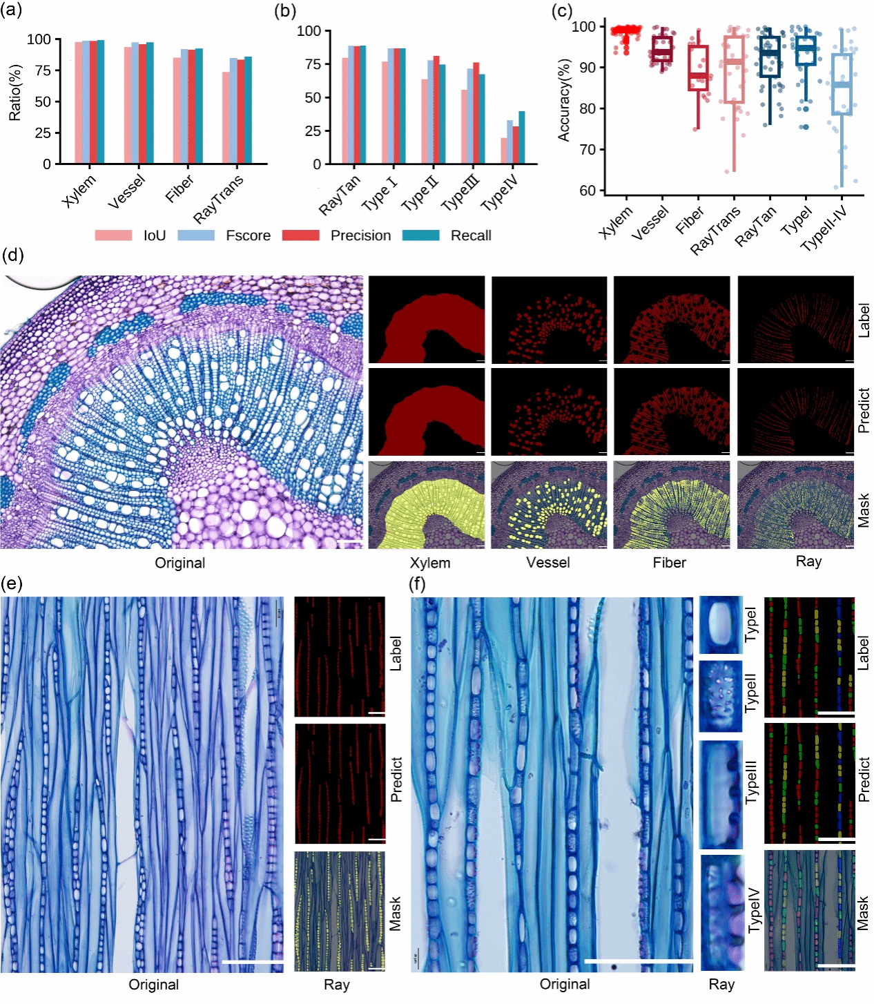
Mask2Former在杨树木质部显微图像上的语义分割性能及代表性叠加可视化结果
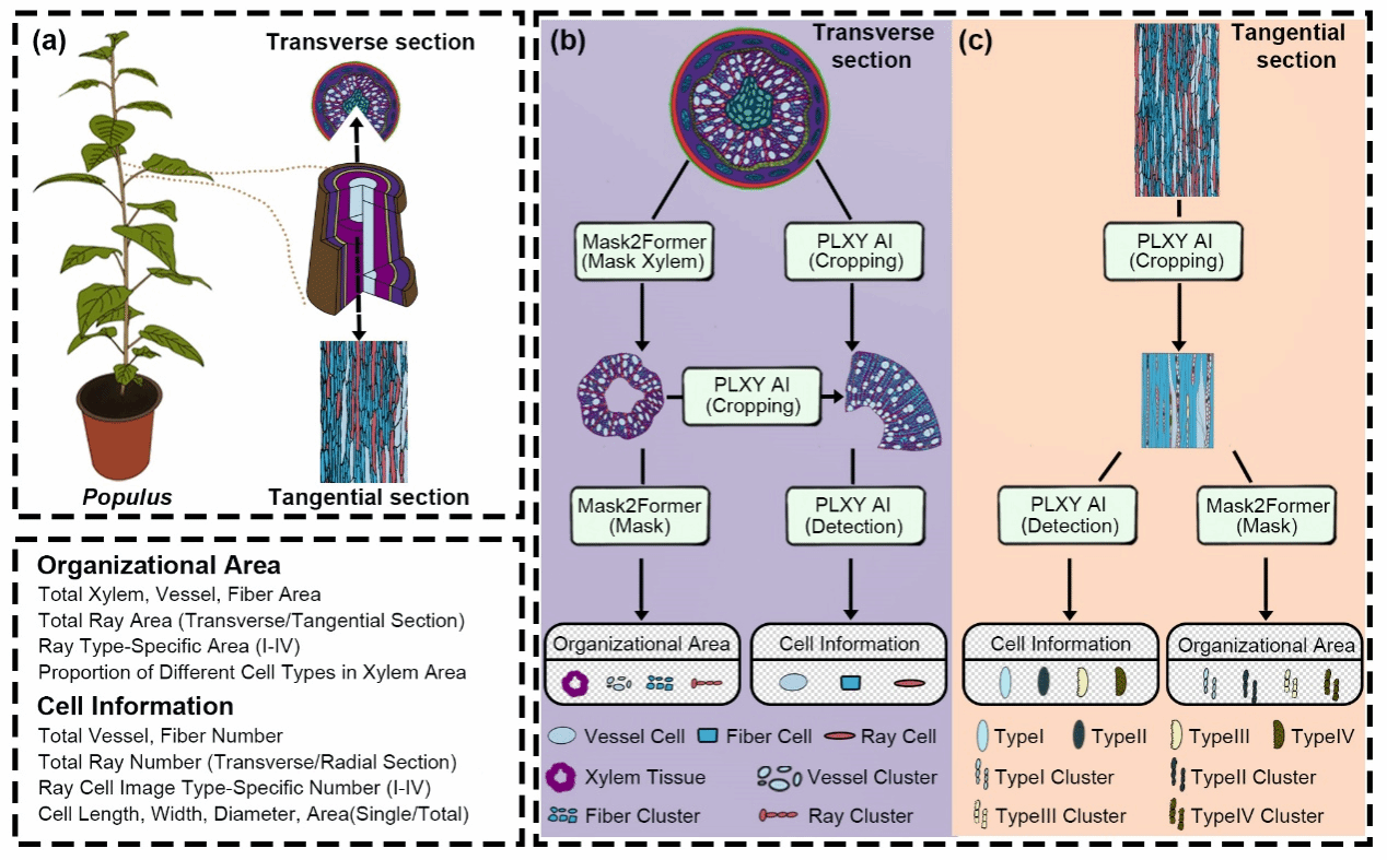
木本植物木质部细胞信息获取的批量化工作流程
我校园艺林学学院已毕业硕士生梅龙威、已毕业博士生戚成和已毕业硕士生裴志阳为该论文的共同第一作者,施雪萍副教授为该论文的通讯作者。华中农业大学郑波教授、包满珠教授、王永健教授和高级工程师傅强,以及浙江农林大学李全梓教授、北京林业大学程瑾副教授参与了本研究。该研究得到了国家重点研究项目和国家自然科学基金的资助。
论文链接:https://academic.oup.com/plphys/advance-article/doi/10.1093/plphys/kiag324/8714972?login=true
【英文摘要】
Xylem is essential for water and nutrient transport, mechanical support, and carbohydrate storage. Identification and quantification of vascular cell types remain manual, time-consuming, and prone to observer bias, limiting throughput and reproducibility. Automated, integrated tools are critical for scaling wood anatomical studies and enabling comparative analyses across taxa. We assembled a poplar xylem dataset of 1,790 microscopy images with 173,434 annotated instances. Using this dataset, we evaluated seven semantic segmentation models and five YOLOv8 detection models across section types for xylem cell recognition and morphometric attribute extraction and adopted a “Segmentation-then-Detection” pipeline to reduce misidentifications in complex backgrounds. Mask2Former achieved the best segmentation performance, covering transverse sections (whole xylem, vessels, fibers, rays) and tangential sections (rays and four ray cell image types). YOLOv8x and YOLOv8m performed consistently for object detection and morphometrics, and the PLXY-AI toolkit was accordingly developed based on YOLOv8 architecture. The combined pipeline markedly improved fiber identification in challenging images. In a generalization test, 34 of 42 woody angiosperms (81.0%) met > 90% accuracy for identifying all cell types. The workflow and PLXY-AI toolkit enable automated identification and quantification of vessels, fibers, and rays, extracting size and area while substantially reducing manual workload and observer bias. Per-image processing time averages < 1 s. Designed for batch analysis, the pipeline minimizes operational complexity and integrates easily into existing laboratory and computational environments. With a user-friendly graphical interface, this framework supports high-throughput analysis of vascular tissue structure and function across multiple tree species.


 最近新闻
最近新闻